After Folloni et al., 2019
Metabolic and Functional Effects of Transcranial Ultrasonic Stimulation (TUS)
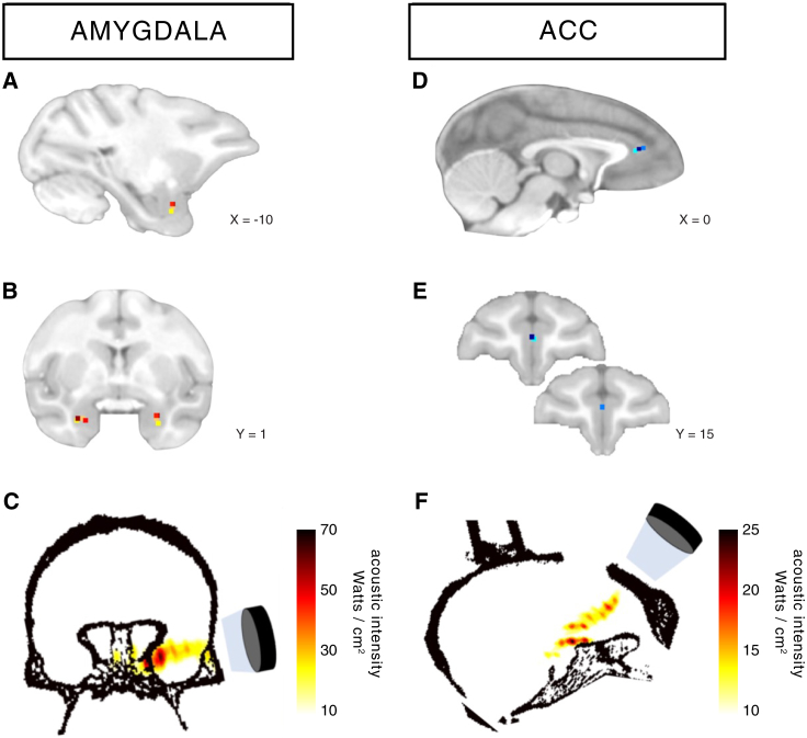
In primates, transcranial ultrasonic stimulation is a preferred method for non-invasive yet reversible disruption of brain activity (Pouget et al., 2020). However, the short-term effects of these disruptions involve mechanisms that are not well understood. While these effects have been studied using functional MRI (fMRI) through functional connectivity (Folloni et al., 2019), the mechanistic effects remain poorly understood.
In this project conducted at the SBRI (Bron, France), we propose to investigate metabolic changes in the area stimulated by TUS using Magnetic Resonance Spectroscopy (MRS) with a non-edited SPECIAL sequence (SPin-Echo full-intensity aCquIred locALized spectroscopy) to better understand the mechanisms induced by these non-invasive stimulations.

"Exploration and Exploitation of Social Scenes in PNH: Awake fMRI
Understanding and integrating social information primarily involves exploring scenes with social actors.
In a task exploring social and non-social scenes through eye tracking, we measure brain activity using fMRI to analyze how social context is interpreted by the prefrontal centers of the cortex, such as the dorsolateral prefrontal cortex (DLPFC) and the cingulate cortex.
This study is part of a larger research project that includes ultrasonic functional imaging and electrophysiology (in other PNH) to better understand the propagation and integration of social stimuli in the primate cortex.
After Abell et al., 2000
Theory of Mind and Metacognition: Macaca mulatta vs. Callithrix jacchus
Theory of Mind (ToM) is the ability to infer emotional states in oneself or others. It develops in humans from around 12 months to 3 to 5 years of age and is an important but non-essential component of human language.
Often mistakenly thought to be a human-specific trait, ToM has been demonstrated in several species, ranging from bonobos to dogs, including marmosets, a New World primate species that lives in large social groups (Dureux et al., 2023).
In this study, we conduct eye-tracking and brain activity measurements using fMRI to compare rhesus macaques and marmosets in order to understand the cortical structures and networks responsible for ToM.
After Claron et al., 2023
Encoding of Social Information by the Medial Cingulate Cortex and Dorsolateral Prefrontal Cortex: A Bimodal Study in Electrophysiology and Ultrasonic Functional Imaging
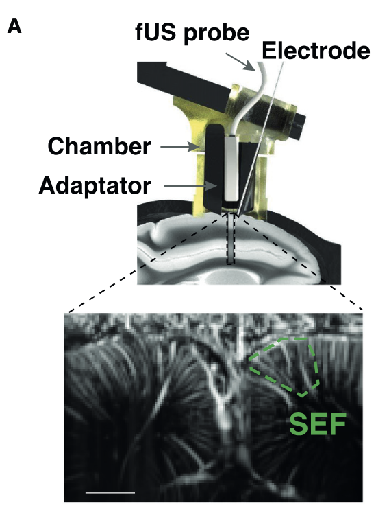
Ultrasonic functional imaging (FUS) offers the advantage of excellent spatial resolution (100µm x 100µm) and good temporal resolution (2.5Hz), albeit at the cost of only allowing imaging of a single plane with an average thickness of 400 µm. Electrophysiology, on the other hand, directly measures neuronal activity within a very limited field of view around the electrode.
In a previous publication (Claron, Provensal et al., 2023), we linked neuronal activity to Cerebral Blood Volume (CBV) activity measured by FUS. In this project, we compare vascular and neuronal activity during the viewing of social scenes to understand the propagation and connection between different brain areas, such as the DLPFC and the MCC, during the extraction of social information.
This will allow us to better understand the dynamics between different parts of the prefrontal cortex during the exploration of complex social scenes.